Our Capabilities
We’re committed to providing specialist ear, nose, throat services, head and neck assessment, and surgical services. Click the below links to find out more about what we do.
- Sleep
Apnoea - Simple snoring in
adults/children - Wax and debris
in the ears - Otitis media
(middle ear infection) - Otitis Externa (External Auditory Canal Infection)
- Perforated
Eardrums - Tonsillitis
- Sinusitis /
Rhinosinusitis - Globus Pharyngeus / Laryngopharyngeal Reflux (Globus/Reflux)
- TMJ
- Tinnitus /
Hyperacusis - Parathyroid
Disorders - Goitre
- Head And Neck
Cancer
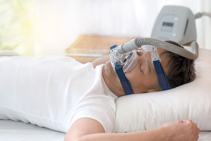
Sleep Apnoea and Snoring
Children who snore often have other stigmata of sleep apnoea, including restless, sweaty, disturbed sleep and are unrefreshed in the morning. They have headaches, and poor concentration and even reduced IQ. It should be noted that snoring is a sign of airway obstruction, usually partial, and is not “normal” or a variation of normality. “Adenotonsillectomy” removal of adenoids and tonsils, can be curative in 80-90% of cases but there are sometimes other contributors, including abnormal facial skeleton or allergic nose (“hay fever”). Surgery is generally very safe, but Prof MacKay and Dr Pearson will detail all of the risks and benefits so that parents are fully aware.

Simple snoring in adults / children
Snoring is also abnormal in adulthood. When complete cessation of breathing (apnoea) or significant oxygen “drop” (hypopnoea) occurs, the condition of Obstructive Sleep Apnoea (OSA) may be diagnosed. The more severe forms of OSA can confer significant risks in terms of the heart, blood pressure, blood vessels etc. Two other common effects are excessive tiredness and partner/family disruption/marital dissatisfaction. Medical, device and surgical treatments might be included.
Prof MacKay is highly credentialed with respect to surgical and multidisciplinary treatment options for snoring and OSA. Please attend appointments with any scans, sleep studies, trials of treatment etc that you may have or previously tried.
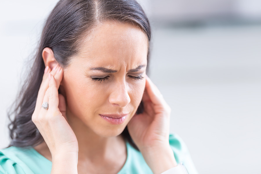
Wax / debris in the ears
Cerumen (“wax”) is a normal component of the ear canal. It is designed as a natural cleaning mechanism. Problems may occur with:
- Over production/excess wax
- Cotton bud use
- Hearing aid use
- Bony ear canal overgrowth (“exostoses”)
- Syringing/ear candle wax/inappropriate or overuse of local treatments Most common problems with wax, debris and foreign bodies can be dealt with safely and effectively in the rooms at Illawarra ENT Head & Neck Clinic. Occasionally, general anaesthesia or sedation may be required.

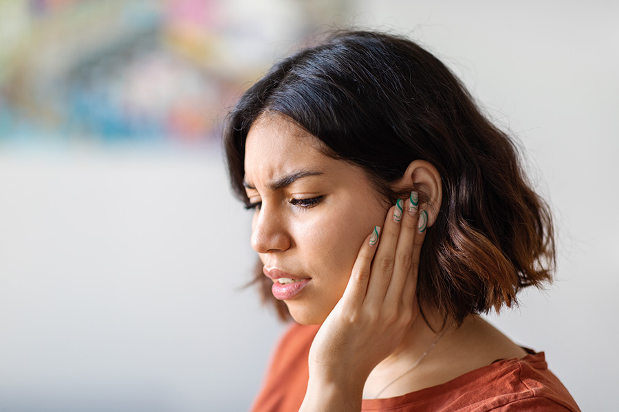
Otitis media (middle ear infection)
Middle ear infections may be acute, chronic (as in “glue ear”) or recurrent acute. At the worse end of the spectrum, surgical treatment ranging from myringotomy (“incision/draining via eardrum”), grommets (small tube that sits across eardrum to equalise pressure) to more complex operations such as incisions behind the ear and drilling of mastoid bone, may be needed. Usually prior to seeing Prof MacKay or Dr Pearson, medical treatments such as antibiotics, steroids, nasal sprays etc have been tried by your GP. Problems with the middle ear should not be ignored, particularly in cases of significant pain, hearing loss or complications (e.g. weakness of the face etc).
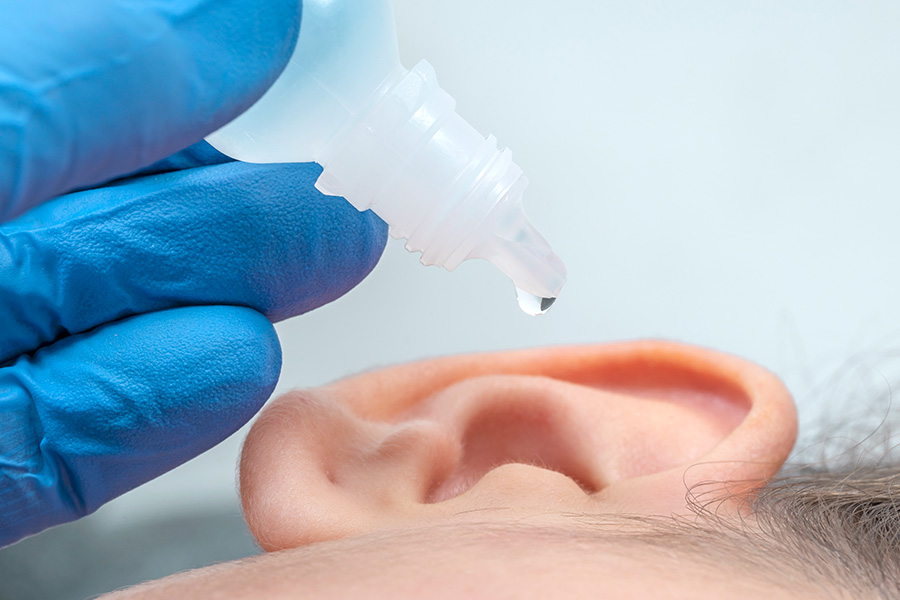
Otitis Externa (External Auditory Canal Infection)
Infections of the ear canal/outer ear usually occur due to cotton bud use, water exposure (swimming, showering, having your hair washed at the hairdresser, etc), hearing aid use, scaly skin (dermatitis) around the outer ear or hairline or overuse of some types of drops into the ear. Infections may be bacterial or fungal. Treatments include antibiotic drops, antifungal drops and ear toilets (cleaning of the ears, preferentially under the microscope). Some patients may require a “wick” in the ear canal, as a delivery system for treatments. Occasionally, in hospital treatment is necessary, and greater concern is always present for patients with suppressed immune systems (e.g. diabetes, cancer, medications that decrease immunity, HIV AIDS, etc).

Perforated Eardrums
A hole in the eardrum (perforated drum or perforated tympanic membrane) usually occurs as a result of infection or trauma. Infective perforations are most common in younger children. 80-90% of perforations will heal/self-resolve, depending on their features, requiring surveillance but no further treatments.
Perforations with infective discharge often require topical drops or cleaning. Avoidance of water exposure is paramount. Some perforations require surgery (myringoplasty) to graft (“heal”) the eardrum. The main purpose for this is to “waterproof” the ear.
Tonsillitis
Tonsillitis refers to inflammation of the palatine tonsils, which are situated in the mouth (at the junction of the “oral cavity” and “oropharynx”). Infective inflammation can be viral, including glandular fever, or bacterial. Your Doctor may prescribe antibiotics and even brief courses of steroids. NH+MRC Guidelines recommend removal of the tonsils if you are getting:
- Seven episodes in one year, or
- five episodes in consecutive years, or
- three episodes a year for three years in a row.
This is especially true if tonsillitis causes missed school, university or work. There are, however, other reasons for removing the tonsils, particularly for snoring or sleep apnoea and when suspicion of cancer/lymphoma exists.


Sinusitis / Rhinosinusitis
Inflammation of the nose and sinuses is termed “rhinosinusitis”. It can occur acutely or chronically or on a recurrent acute basis. In some patients, inflammation is confined more to the nose, “rhinitis” (e.g. allergic – as in hay fever or environmental – as in smokers). Treatments can be derived from a combination of 4 groups:
- Avoidance/reduction (e.g. smoking, house dustmite)
- Medical (e.g. sprays, etc)
- Surgery
- Immunotherapy (e.g. “under the tongue” or injections)
Globus Pharyngeus / Laryngopharyngeal Reflux (Globus/Reflux)
“Globus/Reflux” is one of the commonest clinical presentations to Ear, Nose & Throat Surgeons. It is characterised by a sensation of “sticking”, “mucus” or “furball” in the back of the throat. Patients usually clear their throat excessively, and in two thirds of cases have heartburn or indigestion symptoms. It is essential in cases of prolonged symptoms to exclude more sinister pathology such as cancer, although this is rare. Treatment usually involves a camera examination of the upper airway and digestive tract to confirm the absence of sinister pathology, avoidance of excess throat clearing, drinking frequent sips of water, dietary modifications (especially reducing caffeine, carbonated drinks, spicy foods and alcohol) and anti-reflux medication. Rarely further evaluation with Barium swallow studies or evaluation under general anaesthetic is required.

TMJ
Temporomandibular (jaw) joint dysfunction is very common and symptoms often “mimic” those of ear disease. See our comprehensive patient handout.
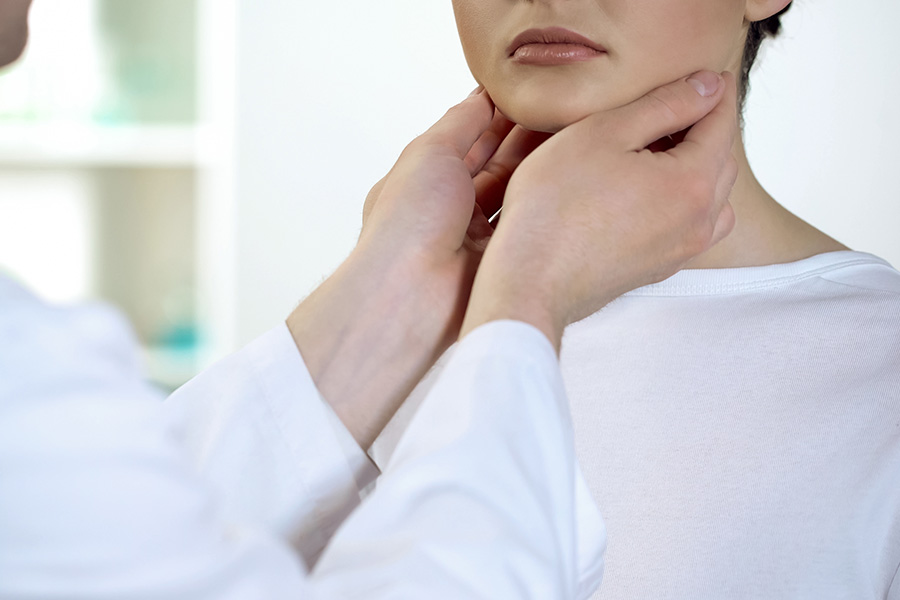
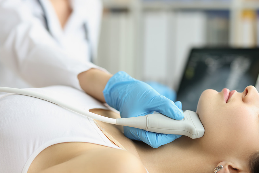
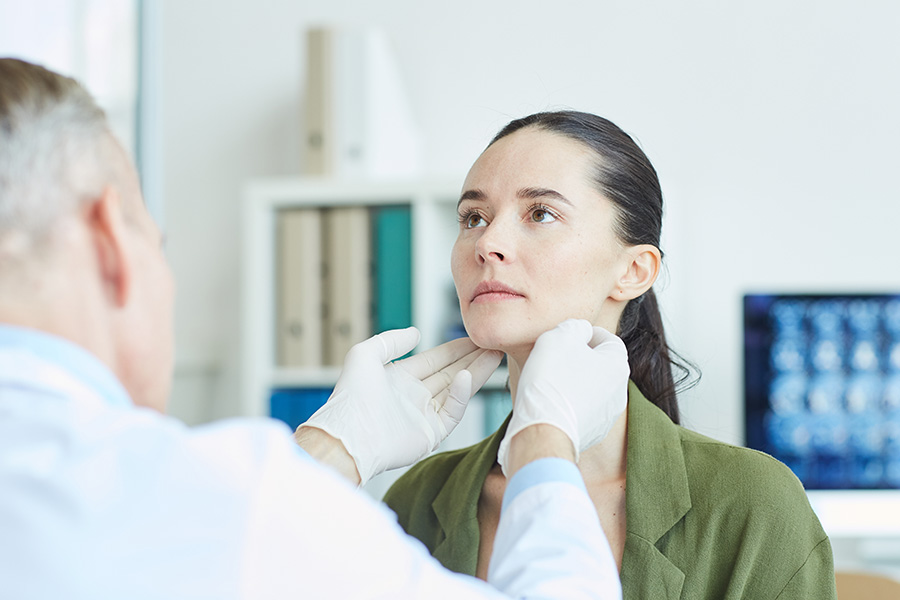
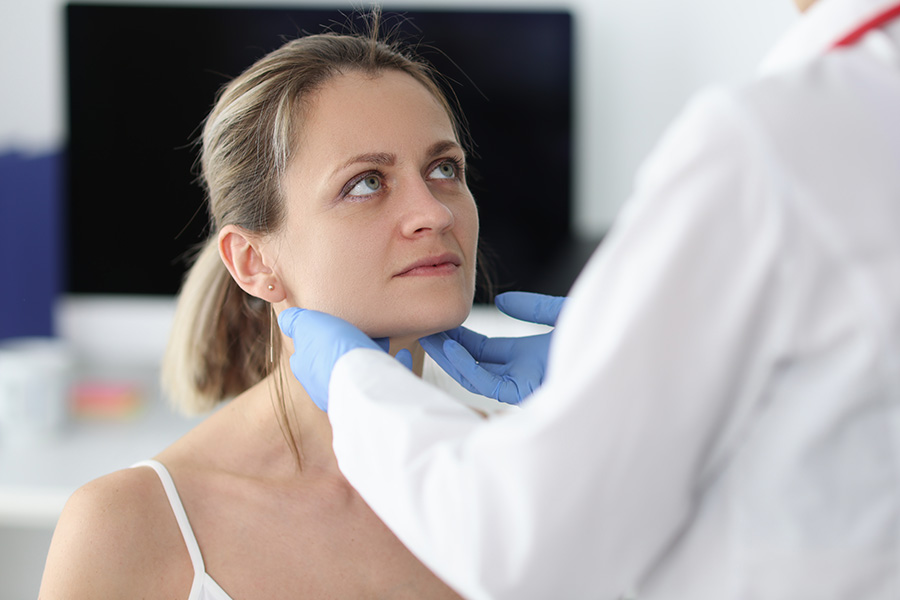
Parathyroid Disorders
- Parathyroid glands are located in the neck or adjacent regions
- disorders affecting parathyroid function alter calcium metabolism
- typically present with bone, joint pain and other systemic complaints
Primary hyperparathyroidism
- usually caused by adenoma – benign tumour of parathyroid gland
- less commonly caused by hyperplasia of all glands
- rarely caused by cancer of parathyroid gland
- treated with surgical exploration of the neck and removal
Secondary hyperparathyroidism
- caused by chronic renal failure
- requires neck exploration with autotransplantation for treatment
Tertiary hyperparathyroidism
- rare condition seen in renal transplant patients
Goitre
Describes enlargement of the thyroid gland – nodular or diffuse
Incidence
- nodules seen in more than 33% of population
- male to female ratio 1:4
Causes include
- iodine deficiency
- autoimmune disease
- Hashimoto, Graves, postpartum
- hereditary defects in thyroid hormone synthesis
- infection
- medication – lithium, iodine excess
- malignancy
Presentation
- incidental swelling in neck without symptoms, or on medical imaging
- local compressive symptoms
- breathing difficulties – lying flat or arms raised
- swallowing difficulties
- voice change – nerve injury
- light headedness with raising arms such as hanging washing
- pain related to rapid enlargement
- bleed into nodule
- inflammation
- cancer – rarely
- endocrine disorder
Assessment
- neck exam – size of goitre and signs of compression, lymph nodes
- voice assessment – video laryngoscopy
Investigations
- blood tests – thyroid function and antibodies, thyroglobulin
- ultrasound with or without fine needle biopsy
- CT scan – for compressive goitre or cancer staging
- nuclear scan – functional assessment of gland / nodules
Medical treatment
- surveillance – small, normal function, benign
- hormone suppression – controversial in benign, euthyroid goitre
Surgery
- compressive goitre
- diagnostic – exclude malignancy
- malignancy
- autoimmune – thyrotoxicosis with systemic complications
Parotid Disease
- The parotid gland is the largest salivary gland – bilateral locations
- located superficial and behind the lower jaw next to the ear
- nerve to facial muscles passes through the gland
Common conditions
- infections
- viral – mumps, other respiratory viruses
- bacterial – usually secondary to dehydration or stones
- rarely cause abscess needing drainage
stones
- obstruct saliva flow causing pain and swelling
- may require surgery to relieve obstruction
tumours
- benign tumours account for 80% of parotid masses
- malignant tumours account for 20%
- metastatic skin cancer is the most common cancer
- surgery is treatment of choice with facial nerve preservation
Head And Neck Cancer
- encompass sinonasal, oral cavity, larynx, and pharynx (throat)
- arise from the lining (mucosa) of the head and neck
- smoking and excess alcohol intake are most common risk factors
- present with symptoms related to the affected site
Assessment
- thorough history of symptoms and risk factors
- complete physical examination of head and neck
- medical imaging – usually CT scan and/or MRI
- tissue biopsy – may require general anaesthetic
- PET scan – nuclear scan part of tumour staging
Management
- discussion at multidisciplinary clinic
- treatment individualised to specific condition
- surgical management combines tumour removal and reconstruction
- radiotherapy / chemotherapy used in particular circumstances
- surveillance after treatment for at least 5 years